The ability of tissues in our body to renew themselves significantly declines with age, imposing the risk for inflammation and cancer. Adult stem cells continuously renew the tissue cells by signalling with their neighbouring niche cells and by relying on their secretory endomembrane system to transport these signals. In the Scharaw lab, we study endomembrane organelle organisation in multicellular stem cell systems, aiming to understand how organelle organisation in stem cells drives tissue renewal and changes with age.
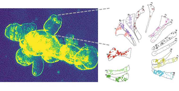
To address these fundamental questions, we use the small intestine as a model, which is marked by unprecedented stem cell-mediated tissue renewal rates, which decline as we age. 3D intestinal organoid cultures allow us to do cell biology in multicellular systems and to apply cross-scale approaches. Specifically, we use ultra-structure volume EM imaging, live-cell transport assays in organoids and mouse genetics.
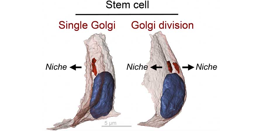
Our group recently discovered that stem cells split and polarise their endomembrane Golgi organelle. This splitting ensures polarised transport of stem cell signals customised towards their niche cells. We found that with age, the Golgi splitting in stem cells is compromised and correlated with declined tissue renewal. Our findings provide a new paradigm, by which stem cells customize their Golgi organelle arrangement for optimized communication with their surrounding in healthy tissue renewal. We aim to unravel the mechanistic principles and functional relevance of endomembrane organelle organisations in stem cell-driven tissue aging. In particular we address:
Our vision is to uncover the principles of endomembrane organelle organisation in stem cell function during health and age-associated diseases.
For further information visit our lab homepage at https://scharawlab.org


Fig. 1. Finding that stem cells split and direct Golgi divisions towards lateral surface contacts with niche cells. Left, volume EM run through an intestinal crypt. Middle, newly discovered stem cell Golgi splitting and arrangement for optimized communication with their surrounding niche cells. Stem cells have one Golgi when in contact with a single niche cell and multiple Golgi’s when surrounded by multiple niche cells. Volume EM retrieved models, Nuclei in blue, cell surface in grey, Golgi in red. Right, newly developed transport assays in intestinal organoids allow signal traffic tracking of single organoid cells using live-cell microscopy.