New research from Barcelona and Dresden: Glycolysis — the process of converting sugar into energy — plays a key role in early development.
More than fuel: Glycolysis doesn’t just power cells — it helps steer them toward specific tissue types at critical moments in development.
Better embryo models: Stem-cell–based embryo models that rely on glycolysis form structures more similar to natural embryos.
Predict and control development in a dish: These findings improve our ability to predict and control how stem-cell-based embryo models will develop, unlocking further potential for biological discovery, disease modelling, and drug toxicity testing.
----------
Glycolysis is an ancient metabolic activity. It consists of a set of reactions that convert glucose into energy. This central process allows cells to grow, divide, and stay alive. It has accompanied life since its origin, from single cells to complex organisms like mammals. Scientists have extensively studied the role of metabolism in individual cells to understand how it influences their energetic state, but little has been studied about the effect of glycolysis on the decisions that cells or groups of cells make.
Now, researchers at EMBL Barcelona, Spain, and at the Max Planck Institute of Molecular Cell Biology and Genetics (MPI-CBG) in Dresden, Germany, uncover the instructive potential of glycolysis. They show that rather than exclusively providing energy to the cell, glycolysis is able to control, at different stages of early embryonic development, cell fate decisions and the end-state appearance of stem cell-based embryo models.
In two back-to-back publications in the journal Cell Stem Cell, researchers from the groups of Vikas Trivedi at EMBL Barcelona and Jesse Veenvliet at the MPI-CBG have used gastruloids and trunk-like structures, in vitro stem cell-based embryo models composed of mouse embryonic stem cells, to study the early steps in the formation of the body plan—a process that lays the foundation for future organ development. Kristina Stapornwongkul, postdoc in the Trivedi group and incoming group leader at the Institute of Molecular Biotechnology (IMBA) in Vienna, Austria, from September 2025, studied the role of glycolysis by changing the glucose concentration in the media where cells live and feed. Alba Villaronga-Luque and Ryan Savill, both doctoral students in the Veenvliet group, studied why some trunk-like structures look more like the natural embryo than others by using machine learning to integrate imaging data with profiles of active genes and metabolites over time and found a critical role for glycolysis.
Stapornwongkul realised that blocking glycolysis disrupted the formation of two important tissue types: mesoderm – which later develops into muscles, bones, or blood – and endoderm – which gives rise to organs like the liver or the lungs. Instead, more cells decided to turn into ectoderm tissue, the type that eventually give rises to our nervous system. This study shows that glycolysis helps activate key signalling pathways (Wnt, Nodal, and Fgf) that guide cells towards mesoderm and endoderm fates. When glycolysis was blocked, the signals weakened and cells developed into ectoderm cells. However, when these signals were artificially boosted, normal cell fate decisions were restored, even without glycolysis taking place. This highlights the crucial role of metabolism as an upstream regulator/activator of specific signalling pathways that influence cellular decisions. Being able to control cell fate through altering media composition means that one could also direct cell differentiation towards the tissue type one is interested in.
“What was most surprising to me was this clear dual role of glycolysis: its bioenergetic function important for growth and its signalling function crucial for cell fate decisions. When we inhibited glycolysis, we clearly saw the loss of the endoderm and mesoderm, but we were able to rescue these cell types by activating the signalling pathways, even in the absence of glycolysis, meaning without restoring growth. This shows that we can decouple glycolysis’s bioenergetic role from its role as an upstream signalling regulator, underlining the existence of two distinct functions during early development,” Stapornwongkul said.

On the left: Glycolysis allows stem cell-based embryo-like models to develop the three germ layers that give rise to many different cell types: ectoderm (magenta), mesoderm (green) and endoderm (yellow). On the right: If glycolysis is inhibited, only ectodermal cells can develop. © Kristina Stapornwongkul
“The exciting thing about Kristina’s work is the hierarchical relationship between metabolism and signalling, at least in the earliest stages of organismal development. This result contributes to an emerging perspective on the relationship between metabolism and patterning, a subject of interest to other labs within EMBL and beyond. From an evolutionary perspective, this is exciting because metabolism predates signalling: even single-cell organisms rely on metabolism, while signalling emerged later in evolution. This has sparked my curiosity about the role of metabolism in the origin of multicellularity. This study marks the beginning of an exciting new direction for my group,” Trivedi said.
Villaronga-Luque and Savill found that early changes in metabolism cause differences in the end-state appearance of trunk-like structures, a stem-cell-based model of embryonic trunk development that forms the tissues giving rise to the spine (mesoderm) and the spinal cord (ectoderm). These structures make it possible to study mammalian embryo development, which is otherwise hidden in the uterus of the mothers, in large numbers of samples without the need for animal experiments. Although many characteristics of these stem cell-based embryo models are similar to those of an embryo, they are unable to develop into fully functional organisms.
A major hurdle for their widespread use is that they are much more variable than the embryo: even when grown under identical conditions, some stem cell clumps develop into structures very similar to the embryo, whereas others don’t. Such variability makes it harder to use them for research purposes that require a highly reproducible baseline, such as disease modelling or toxicity studies. Villaronga-Luque and Savill observed that specifically, the balance between two different processes that produce energy—glycolysis and oxidative phosphorylation—affects the variability of the stem cell-based embryo models. With glycolysis, cells make energy by breaking down glucose. The trunk-like structures with a sort of ‘sweet tooth,’ depending more on glycolysis to obtain their energy by breaking down sugar, develop the most similarly to an embryo, whereas the ones that lacked the ‘sweet tooth’ mostly formed ectoderm. Like Stapornwongkul, they found that glycolysis activates signalling pathways like Wnt that influence cellular decisions and, eventually, how much the structures resemble the embryo. Finally, they showed that boosting glycolysis with drugs improved the appearance of the trunk-like structures.

Collage of stem-cell-based embryo models of the embryonic trunk. Those in the upper row have a ’sweet tooth’, depending more on glycolysis to obtain their energy and resulting in a more embryo-like appearance. In contrast, those in the bottom row have less of a ’sweet tooth’, which results in them producing too much neural tissue, and looking less like an embryonic trunk. © Alba Villaronga-Luque, Ryan G Savill et. al / MPI-CBG
“To uncover the reason for this variability in stem cell-based embryo models, you need to have measures early in the development to see what goes wrong. However, such measurements typically destroy the sample, such that we don’t know if it will develop into a successful structure or not. This is the challenge we were able to tackle.” Villaronga-Luque said.
“By combining quantitative imaging analysis with machine learning, we found key characteristics of the structures that can predict how their development will turn out. With this predictive power in hand, we could then investigate the expression profiles of structures for which the end state would otherwise be unknown,” said Savill.
“By being able to predict the future appearance of a stem cell-based embryo model, our team could show that the early metabolic state controls how much the model looks like the embryo, which can be regulated by altering the metabolic activity with drugs. Such predictive power for stem cell-based embryo models and also other types of organoids has the potential not only to help to make new fundamental biological discoveries but also to improve stem cell-based embryo models for applications that require high reproducibility, including disease modelling, genetic screens, and toxicity studies.” Veenvliet said.
These studies mark the beginning of a new paradigm for developmental and tissue biology. It brings metabolism to the spotlight for studying the early stages of development, giving the research community a new tool to study early cell decisions and embryo development. Excitingly, the two studies show that at different stages of embryogenesis, glycolysis acts through a similar mechanism to ensure proper development of the body plan.
The research in the Veenvliet lab was supported by the European Union’s Horizon Europe Research and Innovation Programme under grant agreement ID: 101071203 (SUMO – Supervised Morphogenesis in Gastruloids), by the Deutsches Zentrum zum Schutz von Versuchstieren (Bf3R) grant (60-0102-01.P589), and by the Stiftung zur Förderung und Erforschung von Ersatz- und Ergänzungsmethoden zur Einschränkung von Tierversuchen (SET) grant.
]]>Chandraniva Guha Ray and Pierre Haas from the Max Planck Institute of Molecular Cell Biology and Genetics (MPI-CBG), the Max Planck Institute for the Physics of Complex Systems (MPIPKS), and the Center for Systems Biology Dresden (CSBD) have now shown that these mechanical instabilities of tissues can be very different from classical instabilities such as the buckling of a sheet of paper under compression: As the compression of a sheet of paper increases, so does the height of the buckled shape. Chandraniva Guha Ray, a doctoral student in the research group of Pierre Haas, explains why this is not the case in a simple theoretical model of a tissue composed of individual cells: “As a tissue folds, the cell sides bend, which causes the top or bottom of the cells to shrink until they cannot bend anymore. This has an unexpected consequence that we called ‘unbuckling’: as the compression increases, the height of the buckled shape can start to decrease.”
Pierre Haas, a research group leader at the MPI-CBG, MPIPKS, and CSBD summarises, “Our calculations prove that this ‘unbuckling’ causes a huge increase of the tissue stiffness. This can be a mechanism for tissue folds to absorb compressive stresses from neighbouring tissues.” One example of a tissue fold that could rely on this mechanism is the so-called cephalic furrow in the fruit fly Drosophila that has recently been studied by the group of Pavel Tomancak at MPI-CBG. Pierre adds, “These minimal mechanical models are useful because they allow us to understand mechanical instabilities in more detail and hence how cell mechanics give rise to biological shape and function at the tissue scale.”
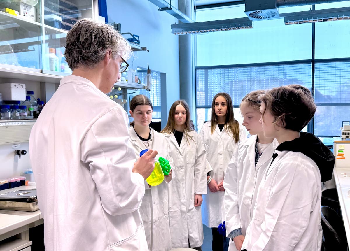
]]>Research group leader Sandra Scharaw and the two postdoctoral researchers Julia Pfanzelter and Émeline Bonsergent discussed their professions and answered questions such as, What is it like to be a scientist? Can you do research while raising a family? Why did you choose to be a scientist? What are your role models? The girls visited the research labs of Eric Geertsma and Mihail Sarov, visited the electron microscopy facility, and enjoyed a panoramic view from the institute roof! The MPI-CBG encourages discussion with interested girls because, even today, female scientists often have a more challenging time in the science world than their male colleagues.
Girls' Day, an initiative of the Federal Ministries for Education and Research (BMBF) and Family Affairs, the Elderly, Women, and Youth (BMFSFJ), is a German-wide campaign that introduces schoolgirls to a variety of careers and activities. Girls are especially encouraged to pursue technical careers in fields where women are still underrepresented, such as "MINT" (mathematics, engineering, natural sciences, and technology).

Tour through the lab of Eric Geertsma © Katrin Boes / MPI-CBG
Early career grants and program grants are awarded to teams of two to four scientists, at any stage of their careers, who embark upon a new collaborative project. HFSP Research Grants support innovative basic research into fundamental biological problems across national and disciplinary boundaries.
"Living systems make guanine crystals for different reasons, like making them to be more sensitive to light (crustaceans) or changing the color of their skin (chameleons). The zebrafish also forms crystals in specialized cells, leading to its brilliant colorful patterns. In our project, my collaborators and I want to find out how this process occurs, considering an essential, but so far unexplored, structure: the lipid membrane that surrounds each crystal”, explains Rita Mateus, who is also an appointed DRESDEN-concept research group leader. Using techniques like CRISPR-Cas9, mass spectrometry, and molecular modeling, the researchers will investigate crystal formation in zebrafish and cell cultures. The results could facilitate the development of therapies to treat diseases where aberrant crystallization occurs, such as kidney stones and gout. The team also plans to contribute new protocols to synthesize organic crystals for a variety of applications in the areas of optics and material science.
The 2025 HFSP Research Grants span the entire spectrum of life science research across the 30 Research Grants and 12 Accelerator Grants that include 104 scientists representing 30 nations. Each grant will last for three years and on average, each award is for $400,000 USD per year. For their awards, the HFSP seeks scientists who form internationally, preferably intercontinentally, collaborative teams, who have not worked together before. In this regard, HFSP fosters frontier research and science diplomacy.
Congratulations to all 2025 winners!
]]>Unlike structured protein regions, where motifs—recurring patterns in protein sequences—are easy to find by using sequence alignments, intrinsically disordered protein regions evolve quickly, and available alignment-based tools are unreliable.
To address this, the research group of Agnes Toth-Petroczy at the Max Planck Institute of Molecular Cell Biology and Genetics (MPI-CBG) in Dresden, Germany, and the Center for Systems Biology Dresden (CSBD) has now developed SHARK-capture, an alignment-free tool for detecting motifs in these challenging disordered regions.
“SHARK-capture compares motifs by using amino acid properties without needing strict rules. The tool can identify conserved motifs more precisely than current algorithms. It means it can better find the exact, often very short region of the sequence that is responsible for a certain function,” explains Chi Fung Willis Chow, postdoctoral researcher in the Toth-Petroczy group and first author of the study.
In collaboration with the group of Simon Alberti at the Biotechnology Center of the TU Dresden, he experimentally characterized a newly detected motif in a protein called Ded1p that the Alberti lab studies. “In my experiments, I changed or deleted the predicted motif, which is only four amino acids long. As a result, the protein had only half of the enzymatic activity, corroborating the functional importance of this short motif,” describes Willis.
Swantje Lenz, a postdoctoral researcher shared between the Toth-Petroczy group and the group of Alexander von Appen at the MPI-CBG, joined the project when the algorithm was developed and was ready to be applied. “By working with SHARK-capture, I was able to give feedback to Willis on how to better score and prioritize motifs. Using SHARK-capture, I identified 10,889 motifs across 2,695 yeast IDRs, providing a valuable resource. I found that many recapitulate already existing experimental data,” says Swantje.
“SHARK-capture is the most precise tool for finding conserved regions in IDRs and is freely available as a Python package. Ultimately, we hope that it will enable the discovery of the sequence determinants that underlie the plethora of functions of disordered regions,” summarizes Agnes.

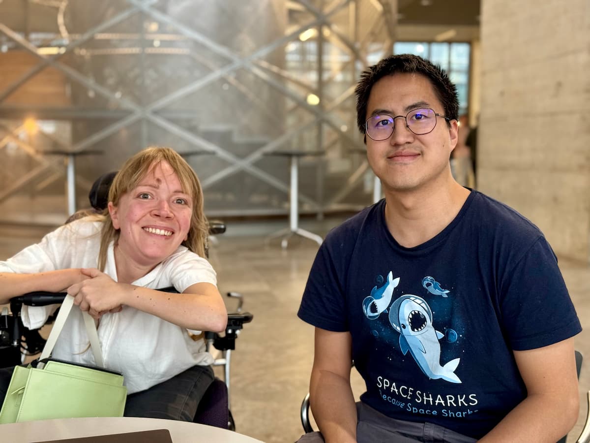
First authors of the study Swantje Lenz (left) and Chi Fung Willis Chow (right). © Katrin Boes / MPI-CBG
Researchers from the Max Planck Institute for the Physics of Complex Systems (MPIPKS) and the Max Planck Institute of Molecular Cell Biology and Genetics (MPI-CBG) have provided new insights into the physical conditions that need to be achieved for enabling an accurate copying process. “Our theory considers both the energy-driven process of making template-based copies and the natural, spontaneous assembly of molecules, as well as the reverse of these processes,” explains Arthur Genthon, the first author of the study pursued together with Frank Jülicher (both MPIPKS), as well as Carl Modes and Stephan Grill (both MPI-CBG). To focus on the core principles of copying, the researchers used a minimal approach based on one-step processes that averages over complex molecular details.
In the minimal setup the researchers considered, they could identify a clear boundary between accurate and inaccurate copy processes. They found that crossing this boundary to achieve accurate copying requires the energy that is spent in the process to surpass a particular threshold. They have thus determined the thermodynamic cost required to achieve precision in replication. The work reveals a cost-accuracy trade-off: using more types of building blocks (monomers) uses less energy but makes copying less accurate. Though inspired by DNA, this framework can apply to any information transfer system, like RNA-to-protein translation.
]]>In her project, Krista wants to compare how the first cell fate decision happens in embryos of different mammalian lineages. During typical early mammalian development, blastomeres (cells produced by the division of the fertilized egg) specialize at the morula stage, becoming progenitors of either the placenta or the fetus. This process, driven by cell position, polarity, and gene activity, has been mostly studied in mice and related mammals. “Afrotherian species like the tenrec, a mammal endemic to Madagascar, develop differently,” says Krista. “Embryos from these species form a central cavity early on without a morula stage. This means that the blastomeres don’t sort into inner or outer positions as they would in a morula. Using this unusual model, I will explore how tenrec embryos make the first cell fate decision without a morula using single-cell RNA sequencing and protein expression analyses. By comparing how this process happens across species, I hope to reveal both shared and unique mechanisms of mammalian development.”
Alexandra’s project will focus on how cell decisions remain stable during development. During cell lineage formation, cells respond to multiple signals to decide on their fate and shape. This process should be robust, even when conditions vary, to reduce the risk of, for example, developmental defects. We know that cells can follow different paths while reaching the same outcome, but relatively little about how much variation in the trajectory can be tolerated or how final decisions are kept on track. Alexandra explains, “I will use stem cell models to follow cell populations as they become more specialized. First, I’ll analyze such fate changes using live imaging and single-cell sequencing. Then, I am planning to test how changing external signals affects the cells’ paths and decisions. In a final step, I want to see if more complex environments can help a cell to stay on course for a specific fate.”
EMBO Postdoctoral Fellowships support excellent postdoctoral researchers throughout Europe and the world. International mobility is a key requirement. The fellowship includes a salary or stipend, a relocation allowance and support for fellows with children. Awardees can attend an EMBO Laboratory Leadership course and become part of the global network of EMBO Fellows.
Congratulations!
]]>The MSCA fellowship is part of Horizon Europe, the European Union’s flagship funding program for research and innovation. The European Commission awarded 417 million euros to 1,696 post-doctoral researchers to work at top universities, research centers, private and public organizations, and small and medium-sized enterprises. The European Research Executive Agency (REA) received 10,360 applications for this call, 16.6 % of which were selected for funding.
“I am interested to study the communication between intestinal stem cells (ISCs) and their neighboring Paneth cells,” says David Grommisch. “As we age, this communication weakens, increasing the risk of inflammation and diseases like cancer. Paneth cells send signals, called ligands, that guide ISCs on how to grow and develop. ISCs receive these signals through receptors on their surface. While we know a lot about how these signals work, we still don’t fully understand how the ligands and receptors are transported inside the cells and delivered to their surfaces. In my project I want to understand this process and how it might help maintaining intestinal health. My plan is to develop a new optogenetic tool in intestinal organoids (mini-gut models) that allows me to track how signals are sent and received in real-time using live imaging. I hope to uncover how aging changes these processes, offering new possibilities for treating age-related intestinal problems.”
MSCA Postdoctoral Fellowships enhance the creative and innovative potential of researchers holding a PhD and wishing to acquire new skills through advanced training and international, interdisciplinary, and inter-sectoral mobility. The funding supports researchers ready to pursue frontier research and innovation projects in Europe and worldwide, including in the non-academic sector.
Congratulations, David!
Press Release of the European Commission: https://marie-sklodowska-curie-actions.ec.europa.eu/news/msca-awards-eu417-million-to-postdoctoral-researchers
]]>Meritxell Huch, Director at the Max Planck Institute of Molecular Cell Biology and Genetics (MPI-CBG), Dresden, Germany, received the prestigious Otto Bayer Award from the Bayer Foundation for her pioneering research on human organoids.
The Executive Director of the Bayer Foundation, Chitkala Kalidas, hosted the event alongside Stefan Oelrich, a member of the Executive Council and member of the Board of Management, and Matthias Berninger, a member of the Board of Trustees, to honor the Bayer Foundation Science Award winners for the Ernst-Ludwig Winnacker Award, the Early Excellence in Science Awards, and the Otto Bayer Award.
Next to Meritxell Huch, four early-career scientists are honored with the Early Excellence in Science Awards. For their exceptional contributions to novel catalytic chemical reactions and their applications in drug discovery, neuroprosthetics and regenerative medicine, reproductive biology, and neuropsychiatric disorders, Richard Liu, Jordan Squair, Ivana Winkler, and Na Cai received the Early Excellence in Science Awards.
The Ernst-Ludwig Winnacker Award 2024 went to Alena Buyx in recognition of her impactful work on public health and ethics policy.
These awards underline the Bayer Foundation's commitment to pioneering research that provides solutions to major health and environmental issues and promotes collaboration between science and practice.
Congratulations to all awardees!
MPI-CBG news on the Otto-Bayer award annuouncement: https://www.mpi-cbg.de/news-outreach/news-media/article/otto-bayer-award-for-meritxell-huch
]]>“As a scientist and lifelong learner, I'm constantly seeking out new techniques and technology to improve workflows and expand knowledge. My background spans many different disciplines, and I thrive on learning from those more advanced than me through constant questioning and conversation,” says Felix Lange. “I am looking forward to working on multidisciplinary levels here at the MPI-CBG.”
Felix Lange studied biochemistry and biomedicine at the Martin Luther University of Halle-Wittenberg and at Leipzig University. For his PhD, he joined the research lab of Stefan Hell at the Max Planck Institute for Multidisciplinary Sciences in Göttingen. He stayed there for his postdoctoral work. During his time in Göttingen, Felix acquired extensive knowledge of the whole portfolio of EM techniques, from the basic ones to cryo-EM.
]]>The workshop, Numerical Nonlinear Algebra in the Real World, featured a broad array of topics through biology, chemistry, and physics with a focus on nonlinear modeling problems and techniques. The talks included local speakers on Active Matter Hydrodynamics (Frank Jülicher, MPI for the Physics of Complex Systems, MPIPKS), Stories of Ecologies (Pierre Haas, MPIPKS and MPI-CBG), and Mathematical Machine Learning (Jiayi Li, MPI-CBG). External presenters included Fast and Flexible Modeling of Chemical Reaction Networks (Torkel Loman, University of Cambridge) and Equilibria in Game Theory (Irem Portakal, MPI for Mathematics in the Sciences).
The communication spaces in MPI-CBG and the Center for Systems Biology Dresden (CSBD) fostered collaborations between early-career PhD students and researchers and faculty, spanning polynomial optimization, tropical geometry, and homotopy continuation. The whiteboards in the atrium facilitated open discussions and new research projects. This open and inclusive environment also allowed us to display mathematical art, for example, a 3D-printed interactive Barth Sextic created by Silviana Amethyst. Conversations continued online through video profiles and vignettes of the talks posted by Anna Frangou on the Math in Science Bluesky account (@math-mpicbg.bsky.social), which spurred engagement with the wider scientific community.
“The workshop was a real pleasure,” said Aida Maraj, a new mathematics group leader here at the CSBD. “It’s always inspiring to meet new collaborators as well as continue older conversations. And it’s wonderful to see so many people contributing to our mission of bringing disciplines together.”
The hub of mathematics at the MPI-CBG is rapidly growing. The research groups colocated at the CSBD plan to host many more research events in the future (workshops, schools, etc.) to close the gap between mathematics, computation, physics, and biology.

3D-printed interactive Barth Sextic created by Silviana Amethyst. ©Katrin Boes / MPI-CBG
]]>The “TUD Young Investigator”scheme aims to counteract the structural disadvantages sometimes experienced by this group of researchers due to their lack of defined status and inadequate or nonexistent connection to a faculty.
Every "TUD Young Investigator" will be paired with a TU Dresden professor as mentor and will be accepted by the faculty as examiner in doctoral procedures, particularly with regard to the dissertations they (co-)supervise. Additionally, the junior group leaders will get the opportunity to take part in teaching and various trainings individually designed to address the needs of academics in this qualification phase.
Congratulations to Aida and Türkü!
]]>Researchers in the group of Agnes Toth-Petroczy at the Max-Planck Institute of Molecular Cell Biology and Genetics (MPI-CBG) and at the Center for Systems Biology Dresden (CSBD) have now developed a machine learning classifier (a type of algorithm) that is less biased towards the proteins with high level of disorder. The classifier PICNIC—Proteins Involved in CoNdensates In Cells—accurately predicts proteins that form condensates by learning the amino acid patterns in protein sequences and structures, along with their intrinsic disorder features. Anna Hadarovich, one of the two lead authors of the publication in Nature Communications and a postdoctoral researcher in the group of Agnes, explains, “We trained the classifier with proteins from human. However, I was positively surprised to see how well the predictions of PICNIC worked on other species that it wasn't trained on. We proved this with previously published experimental data.” Hari Raj Singh, the second lead author and postdoctoral researcher in the group of Anthony Hyman, who is a director at the MPI-CBG, performed the experimental validation of the classifier PICNIC. He says, “We tested 24 proteins predicted to be part of condensates in cells and found the tool to be about 82% accurate, regardless of how much structural disorder the proteins had.”
“We developed a machine-learning tool that can analyze condensate proteins across entire proteomes, the complete set of proteins produced by a cell, in different organisms. PICNIC shows that it can identify general patterns using only protein sequence information and structures derived from it across many different species,” says Agnes Toth-Petroczy, who oversaw the study, and continues, “These results can help us understand how biomolecular condensates have evolved and predict more proteins involved in condensates. This could also help identify protein targets for modifying diseased condensates and aid drug development.” The classifier PICNIC is open-source Python package that is easy to use, so everyone can use it for any protein, synthetic or real from different species.
]]>The research group of Agnes Toth-Petroczy seeks to understand protein sequence space and the collective organization of proteins into biomolecular condensates in the light of evolution and erroneous protein production, i.e. transcription and translation errors that lead to phenotypic mutations. Agnes says, “Proteins are the most intriguing biomolecules of life, driving essential functions. We use an interdisciplinary approach combining computational and experimental biology to advance systems level understanding of proteins and biomolecular condensates in evolution, health and disease. With my membership in the EMBO Young Investigator Programme, I hope to further my research here in Dresden, and I feel very honored to be part of such a vibrant community.”
As part of the Young Investigator Programme, Agnes has access to a lot of networking opportunities for herself and her lab members. The young investigators, who receive an award of 15,000 euros, also benefit from training in laboratory leadership and responsible conduct of research, access to core facilities at EMBL in Heidelberg, Germany, and mentoring by EMBO Members. They can apply for additional grants, for example, for organizing or travelling to conferences.
Of the 27 new EMBO Young Investigators, 14 are female (52%) and 13 are male (48%). They are based in 10 member states of the EMBC, the intergovernmental organization that funds the EMBO Programmes, and Japan. In total, the program received 207 eligible applications, and the success rate was 13%. In the framework of the memorandum of cooperation between EMBO and the Japan Science and Technology Agency (JST), scientists funded by certain programs of JST were eligible to apply to the EMBO Young Investigator Programme for the first time in 2024.
About EMBO
EMBO is an organization of more than 2,100 leading researchers that promotes excellence in the life sciences in Europe and beyond. The major goals of the organization are to support talented researchers at all stages of their careers, stimulate the exchange of scientific information, and help build a research environment where scientists can achieve their best work.
]]>The non-profit Boehringer Ingelheim Foundation is supporting the project with EUR 20 million over a period of ten years, thereby providing half of the EUR 40 million total. The Max Planck Society, TU Dresden and the Free State of Saxony are financing the other half of the project, which aims to facilitate research in the field of biological and biomedical AI. To this end, a new division with two research groups is being established at the Center for Systems Biology Dresden (CSBD), an inter-institutional center jointly run by the Max Planck Institute of Molecular Cell Biology and Genetics (MPI-CBG), the Max Planck Institute for the Physics of Complex Systems (MPIPKS) and TUD Dresden University of Technology.
The new research division will also work in close partnership with the AITHYRA Institute in Vienna, which was established in September 2024 and is also funded by the Boehringer Ingelheim Foundation. DeepMind Professor Michael Bronstein has been recruited as Founding Director for this unique European Institute devoted to artificial intelligence in the field of biomedicine.
BioAI Dresden combines innovative AI methods with knowledge from biochemistry and physics across the entire spectrum of biology, with the aim of making a decisive contribution to a new scientific understanding of our health. The selection and appointment of directors and research group leaders will follow the excellence criteria and procedures of the Max Planck Society.
The combination of biomedicine and artificial intelligence holds enormous potential – potential that can only be realized through comprehensive and interdisciplinary collaboration. This is why BioAI Dresden and the AITHYRA Institute are seeking out partnerships with outstanding research institutions such as the European Molecular Biology Laboratory (EMBL) with its six sites in Europe, EPFL – Swiss Federal Institute of Technology in Lausanne, the University of Oxford, and the Broad Institute in the USA. The new research project will enable Dresden to fully use and develop its potential.
Michael Kretschmer,
Minister President of the Free State of Saxony
“When it comes to medical and biotechnology, Saxony is a world-renowned hub of expertise. In recent years, significant progress has been made in this area thanks to research and development, enabling diseases to be identified and treated more effectively. With this new research program, we are setting the course for the future. Artificial intelligence will increasingly become an integral part of our daily lives in the years to come. We also want to exploit these opportunities for biomedicine. I would like to thank the non-profit Boehringer Ingelheim Foundation, the Max Planck Society and TU Dresden for their tremendous dedication, and I wish the researchers every success.”
Sebastian Gemkow,
Minister of Science of the Free State of Saxony
“With BioAI Dresden, another unique and outstanding field of research is emerging in the science hub of Saxony. The Max Planck Society and TU Dresden are joining forces with the Boehringer Ingelheim Foundation to combine their expertise in biomedicine and artificial intelligence, thus breaking into a new field of research. This has the potential to yield completely new approaches for biological systems across different levels, to explain the underlying principles of how biological systems function, and to predict their response to disruption. As a result, it paves the way for new forms of treatment in medicine and pharmaceutics. I am convinced that this new research division will very quickly gain an international reputation as an institute for cutting-edge research and be perpetuated as a university alliance.”
Christoph Boehringer,
Chairman of the Boehringer Ingelheim Foundation
“The recent Nobel Prize awards have emphasized the huge potential of AI and biomedicine for human health. The non-profit Boehringer Ingelheim Foundation is committed to creating the best possible conditions for independent research in this field in Europe. Our commitment is also intended to inspire the European ideas of scientific freedom and international collaboration. An outstanding collaboration that serves the well-being of all people in Europe and beyond.”
Dr. Stephan Formella,
Managing Director at the Boehringer Ingelheim Foundation
“If we are to establish a relevant European focus from a global perspective in the field of AI and biomedicine, we need strong collaboration between different stakeholders. As a non-profit, independent foundation, we see it as our mission to build bridges between these groups. This means that every site we support can act as an individual pillar in the future, with the resulting bridges creating an even greater impact. In addition to our support in Vienna, we have now also laid the foundations for this in Dresden.”
Prof. Patrick Cramer,
President of the Max Planck Society
“The MPG has long been a European driving force in the field of AI. The journal NATURE lists us in 7th place among the top 10 “Rising Institutions in Artificial Intelligence”. In collaboration with the Austrian Academy of Sciences and with the support of the Boehringer Ingelheim Foundation, we now want to join forces further in this area of research. After all, there are many good reasons not to leave this field to the big tech companies alone. Basic research can and will address questions that profit-driven companies do not take up, but which can be of great benefit to the general public.”
Prof. Ursula Staudinger,
Rector of TUD Dresden University of Technology
“By signing this agreement today, we are building another bridge between disciplines, stakeholders and locations. Thanks to the support from the Boehringer Ingelheim Foundation, the Max Planck Society, and the Free State of Saxony, two new research groups can now be established and an inaugural director appointed at the Center for Systems Biology at TUD, with the aim of developing an innovative branch of research at the interface between AI and biomedicine. This project strengthens the ties between TUD and the Max Planck Institute of Molecular Cell Biology and Genetics, which are jointly conducting research in the CSBD – including in the team of Ivo Sbalzarini, who is also a Professor and Dean at TUD's Faculty of Computer Science. What's more, this project is a good complement to our core research areas in the field of digital sciences. As one of nine national high-performance computing centers and with lighthouses such as CIDS and SCADS.AI, TUD offers excellent infrastructure and paves the way to additional cooperation opportunities.”
Prof. Stephan Grill,
Director of the Max Planck Institute of Molecular Cell Biology and Genetics
“Living systems are incredibly complex. AI will be key to unraveling this complexity and understanding how living systems work. This exciting joint project will allow us to develop a new generation of physics-informed biomedical AI algorithms for identifying the principles and mechanisms that make up living systems. We are therefore ideally positioned here to drive the next revolution in the life sciences.”

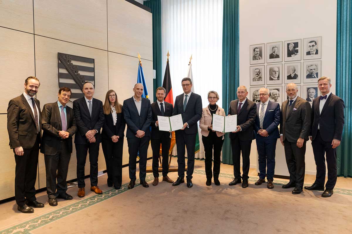
Minister of Science Sebastian Gemkow; Prof. Stefan Bornstein, UKDD; Marc Wittstock, Managing Director BIS; Prof. Heather Harrington, Director MPI-CBG; Prof. Stephan Grill, Director MPI-CBG; Minister President Michael Kretschmer; Prof. Patrick Cramer, President of the MPG; Prof. Ursula Staudinger, Rector of TUD Dresden University of Technology; Dr. Dr. Michel Pairet, Member of the Board of the BIS; Dr. Stephan Formella, Managing Director at the BIS; Christoph Boehringer, Chairman of the BIS; Minister of State Dr. Andreas Handschuh (v.l.). © Pawel Sosnowski
Boehringer Ingelheim Foundation
The Boehringer Ingelheim Foundation is an independent, non-profit organization committed to the promotion of the medical, biological, chemical, and pharmaceutical sciences. It was founded in 1977 by Hubertus Liebrecht, a member of the Boehringer family, co-owners of Boehringer Ingelheim. With its “Plus 3,” “Exploration Grants” and “Rise up!” funding programs, it supports excellent researchers in crucial phases of their careers. It also funds the international Heinrich Wieland Prize and awards for emerging scientific talent, and supports projects at various institutions such as the Institute of Molecular Biology (IMB) in Mainz, the European Molecular Biology Laboratory (EMBL) in Heidelberg, and the AITHYRA Institute in Vienna. https://boehringer-ingelheim-stiftung.de/en/index.html
TUD Dresden University of Technology
As a University of Excellence, TUD Dresden University of Technology is one of the leading and most dynamic research institutions in Germany. With around 8,300 staff and 29,000 students in 17 faculties, it is one of the largest technically-oriented universities in Europe. Founded in 1828, today it is a globally oriented, regionally anchored top university that develops innovative solutions to the world's most pressing issues. In research and teaching, the university unites the natural and engineering sciences with the humanities, social sciences and medicine. This wide range of disciplines is an outstanding feature that facilitates interdisciplinarity and the transfer of science to society.
MPI-CBG
The Max Planck Institute of Molecular Cell Biology and Genetics (MPI-CBG), located in Dresden, is one of more than 80 institutes of the Max Planck Society, an independent, non-profit organization in Germany. 550 curiosity-driven scientists from over 50 countries ask: How do cells form tissues? The basic research programs of the MPI-CBG span multiple scales of magnitude, from molecular assemblies to organelles, cells, tissues, organs, and organisms. The MPI-CBG invests extensively in Services and Facilities to allow research scientists shared access to sophisticated technologies.
“The field of fluorescent microscopy is very multidisciplinary. This multidisciplinarity is what fascinates me about this field. Optical microscopy has the unique ability to look inside living cells and visualize biological processes as they are taking place. The fundamental challenge of optical resolution has been overcome in recent decades; however, these developments have come at the cost of a reduced acquisition speed. In most cases, to such a low level that the methods are limited to fixed samples. My goal at the MPI-CBG is to push the speed of high-resolution microscopy techniques to uncover and understand fundamental biological processes.
Welcome to the institute, Michael!
Michael studied physics at the Technical University of Munich. In 2016, he started his Ph.D. work at the Max Planck Institute for Multidisciplinary Sciences in the research group of Stefan W. Hell with a focus on Nano-Biophotonics. For his Ph.D. thesis he was awarded with the Otto Haxel Award for Physics in 2021 and with the Otto Hahn Medal in 2022. Michael continued his postdoctoral work in the group of Stefan W. Hell until 2024, when he became a research group leader at the MPI-CBG.
]]>Meritxell Huch is receiving the award in recognition for her pioneering research on human organoids. Her work has significantly advanced the use of organoid models in drug discovery, screening, and disease modeling for personalized medicine. Organoids are small, in vitro organ-like structures derived from stem cells. Meritxell and her team have studied the growth and regeneration of animal and human liver and pancreas organoids. Her research is of great importance for the development of new therapies to combat life-threatening cancer in these organs without the need for animal testing. For her scientific work, Meritxell has already received several awards, including the EMBO Young Investigator Award and the German Stem Cell Network Award. In 2023, she was elected to become a member of the European Molecular Biology Organization. Since May 2024, Meritxell Huch has been an honorary professor for stem cell research and tissue regeneration at the Medical Faculty of the TU Dresden.
The Otto Bayer Award is presented alternating with the Hansen Family Award every second year. It recognizes leading scientists working in German-speaking countries for ground-breaking research in chemistry or biochemistry. It was established in 1984 by a provision in the will of Professor Otto Bayer, a former Director of Research at Bayer.
Press Release of the Bayer Foundation: https://www.bayer-foundation.com/winners-announcement-otto-bayer-award-and-early-excellence-science-awards-2024
]]>The research groups of Anne Grapin-Botton at the Max Planck Institute of Molecular Cell Biology and Genetics and of Frank Jülicher and Marko Popović, both at the Max Planck Institute for the Physics of Complex Systems, set out to investigate the rotation of spherical cell clusters. The researchers looked at lab-grown mouse pancreas organoids—models of an organ in three dimensions. Tissue rotation is quite common and is also observed in other organoid systems or in embryos.
“We found that many of these cell clusters rotate continuously, with the direction of rotation sometimes drifting or stopping entirely,” says Tzer Han Tan, one of the three lead authors of the study. He continues, “We asked ourselves why the cell clusters (or organoids) are rotating and reached out to our physicist colleagues Frank Jülicher and Marko Popović.” The researchers built a three-dimensional physical model to understand the collective cell behavior. Those 3D vertex models are usually used to describe other things, but here, the researchers added a polarity vector for cells to the system and set up the model on a round sphere. “By running a simulation and then comparing it with experimental data for rotation speed and the stability of the rotation axis, we were able to show that the interplay of traction forces and cell polarity can explain these rotational behaviors,” say Frank Jülicher and Marko Popović.
Spherical tissue rotates solidly most of the time, like the earth, but the sphere can sometimes transition to a flowing state and spontaneously break symmetry. The collective cell behavior can give rise to this chiral asymmetry in three dimensions. An object or a system can be described as chiral if it is distinguishable from its mirror image. Both rotational motion and turbulent flows are necessary to achieve chiral asymmetry.
Anne Grapin-Botton, one of the three supervising authors, summarizes, “The robust mechanism for chiral symmetry breaking that we have discovered may have implications to understand the development of left-right asymmetry in biological systems. Our results most likely apply to rotating spheres composed of many cell types and in the pancreas may be deployed on other geometries such as tubes. This may be essential for understanding how cells behave on complex three-dimensional surfaces.”
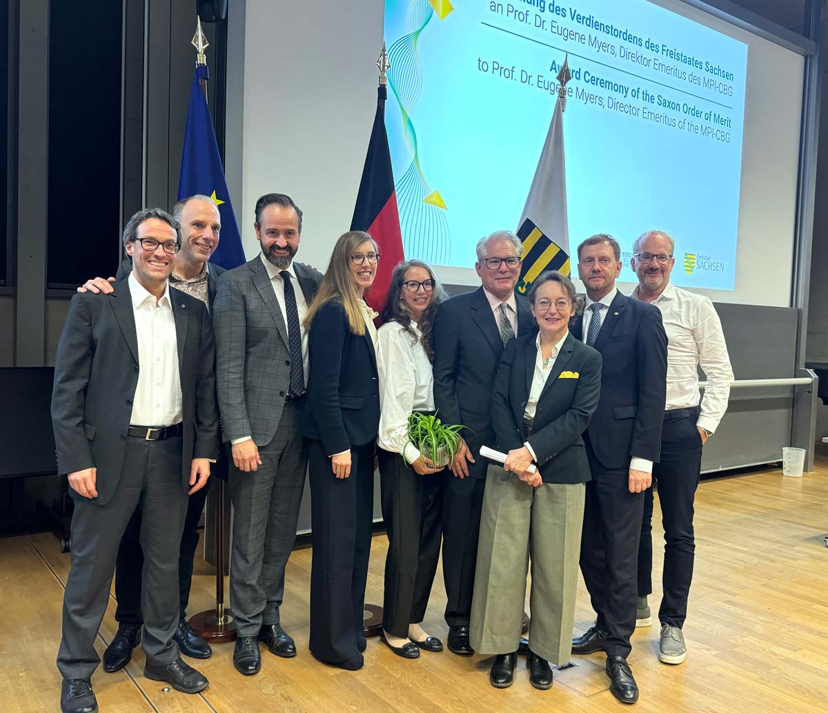
]]>Michael Kretschmer, Minister President of Saxony, presented the award to the 70-year-old at the MPI-CBG during a symposium on October 29.
The mathematician is one of the pioneers in the field of bioinformatics, and his scientific work and the BLAST algorithm, which he co-developed, were crucial to the decoding of the human genome.
In his laudatory speech, Kretschmer also referred to Prof. Myers' special achievements in strengthening Saxony as a center of science and research, and in particular the biotechnology cluster in the Free State of Saxony. “You have made groundbreaking achievements and shown outstanding commitment to Saxony as a center of biotechnology.” All of this is invaluable, he said.
The Saxon Order of Merit is the highest state honor in Saxony. With this award, the Free State honors people who have shown outstanding commitment in the political, economic, cultural, social, societal, or voluntary sectors. The order was established in 1996 and first awarded on October 27, 1997. It can be awarded to individuals from Germany and abroad who have made a particular contribution to the Free State of Saxony and the people who live here. So far, the Saxon Order of Merit has been awarded 402 times.

Prof. Dr. Ivo Sbalzarini, Prof. Dr. Stephan Grill, Saxon Science Minister Sebastian Gemkow, Prof. Dr. Heather Harrington, Daphne Myers, Prof. Dr. Eugene Myers, Dr. Anne Grapin-Botton, Saxon Minister President Michael Kretschmer, and Prof. Dr. Anthony Hyman (from left to right) © Katrin Boes / MPI-CBG
]]>“My work is in an interdisciplinary area called algebraic statistics. Some models arrive from phylogenetics or can be used to model data from biology.” says Aida Maraj. “Mathematics is a new part at the MPI-CBG and CSBD. I see it as a privilege to build new things here together with my colleagues. ”
Welcome to the institute, Aida!
Aida studied mathematics at the University of Tirana in Albania and moved in 2015 to the University of Kentucky, USA, for her PhD in Mathematics with a thesis on Algebraic and Geometric properties of Hierarchical Models. In 2020 she went to Leipzig as a postdoctoral researcher at the Max-Planck-Institute for Mathematics in the Sciences. After a year, she moved back to the USA with postdoctoral positions at the University of Michigan and Harvard University before starting her position as research group leader in Dresden. Her research has been supported with grants by the American Mathematical Society and the National Science Foundation.
]]>